Technology
Stability built into the container.
DuraRad is a thin sol-gel enzyme coating bonded to the inside wall of the vial. As the drug's own radiation splits the water around it into reactive oxygen species, the coating consumes the superoxide and peroxide that reach the wall, holding their level in the solution low, with no change to the drug product.
The container
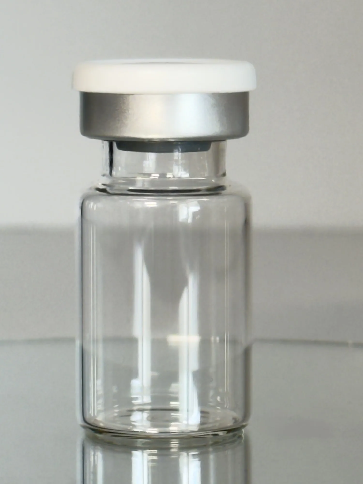
A standard serum vial.
- 6R borosilicate tubular glass
- Standard ISO formats; the same stoppers and seals, sealing against uncoated glass
- Designed to arrive sterile: closed stoppered vials, or unstoppered in a sealed tub
The mechanism
Lower reactive oxygen species at the wall.
DuraRad is a thin sol-gel coating bonded to the interior wall of a standard pharmaceutical vial. It immobilizes two scavenger enzymes, catalase and manganese superoxide dismutase, in a stable, inert silica network.
As radiolysis generates reactive oxygen species in the solution during storage, the wall-bound enzymes continuously clear the superoxide and the peroxide it leaves behind, holding both to a low level over the shelf life and lowering the drug's cumulative oxidative exposure. Radiochemical purity, the share of the dose still in its intact and active form, holds longer. Whether the coating keeps clearing under live decay is what the radioactive validation study is designed to measure.
Because the enzymes are locked into a silica matrix that is bonded to the wall, they act as a fixed feature of the container rather than a formulation additive: present through storage, with no enzyme leaching detected in cold bench challenge testing.
Figure 1 · Radiolytic degradation
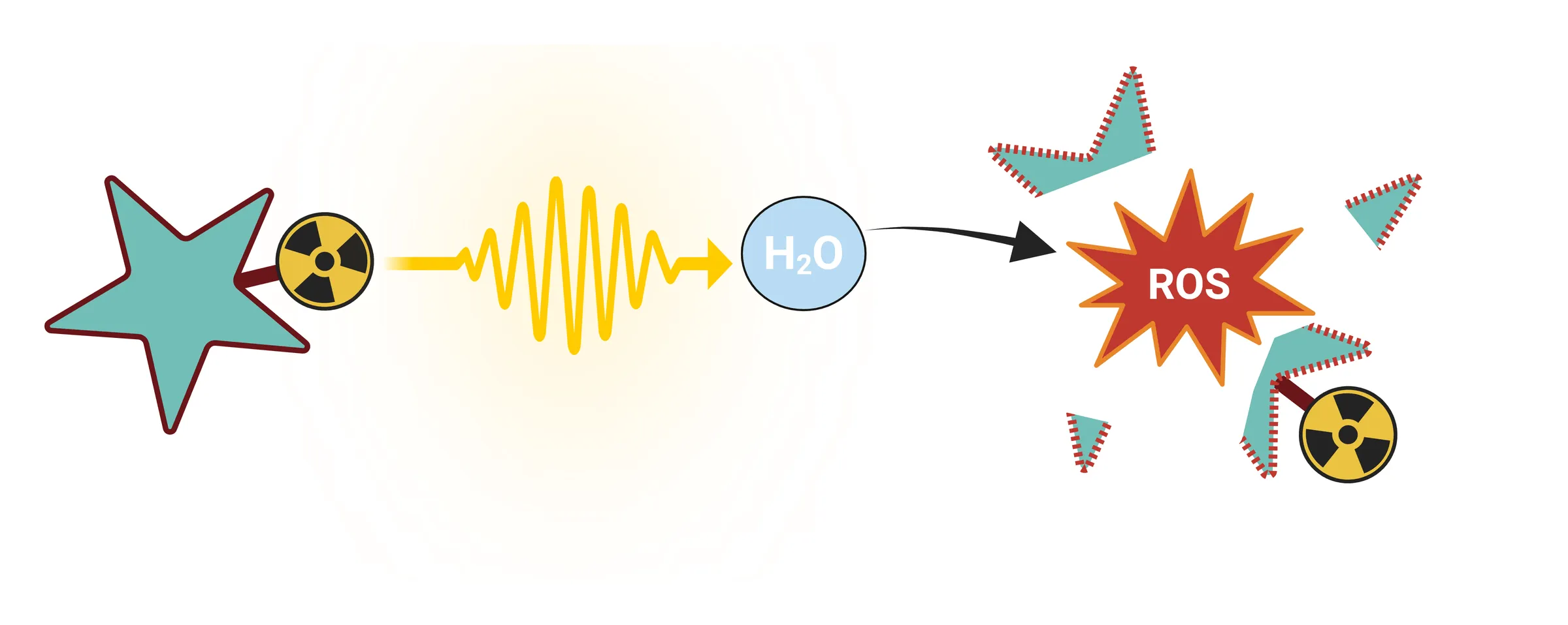
Radiolysis, at the scale of the molecule. Most of the damage is not radiation striking the drug directly. Decay energy is absorbed by the water the drug is dissolved in, splitting it into reactive oxygen species, and those species then oxidize the drug that produced them. That indirect path is what makes the problem addressable at the container instead of in the formulation.
Simplified. Water radiolysis produces a cascade of species; this figure collapses it into a single burst to show the pathway from decay to damage. The individual species, and which of them the coating acts on, are drawn in Figure 2.
Created in BioRender. Wiley, C. (2026) BioRender.com/qkow2ss (opens in a new tab)
Figure 2 · The DuraRad system
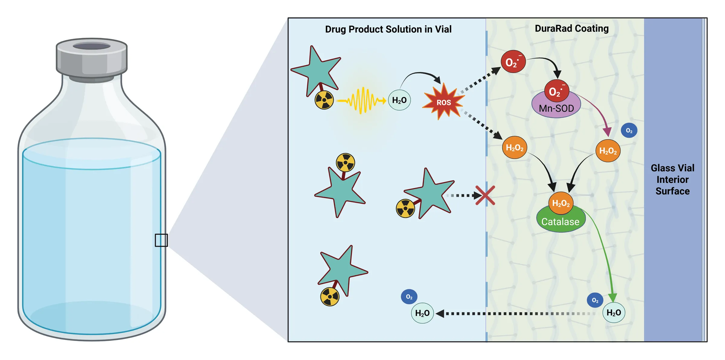
The coated wall, magnified. The panel runs from the drug product solution on the left, through the DuraRad coating, to the glass surface it is bonded to. The superoxide and hydrogen peroxide that reach the coating are consumed there by the immobilized enzymes and leave as water and oxygen, both ordinary constituents of the solution. The red cross is the size argument: the silica network is open to small molecules and closed to large ones, so the radiolysis products diffuse in and the intact radioligand is too large to follow. And because these enzymes are catalysts rather than sacrificial scavengers, a site is not used up when it acts.
- The drug carries its own radiation source. Decay energy leaves the radionuclide in every direction, into the solution around it.
- Where that energy passes through water it splits it. The fastest radicals react within nanometers of where they form; what persists and spreads through the liquid is chiefly superoxide and hydrogen peroxide.
- Those species diffuse at random. The ones that reach the coated wall pass into the silica network, where the enzymes are held.
- Mn SOD converts two superoxide radicals into hydrogen peroxide and oxygen. Catalase takes two of those peroxides and returns water and oxygen.
- Water and oxygen diffuse back into the solution, and each enzyme site is left free to clear again; a site is not consumed by acting.
- The radioligand is too large to follow. The network is sized to admit small molecules like peroxide, not a whole drug conjugate, and the same pore structure is what holds the enzymes in place.
Illustrative schematic, not to scale: the coating is a thin film on the glass surface, drawn wide enough here to show what happens inside it. Water radiolysis yields more than the two species drawn, among them hydroxyl radical, hydrated electrons and hydrogen atoms, with singlet oxygen arising in secondary reactions; Mn SOD and catalase act on superoxide and hydrogen peroxide, and hydroxyl radical is not an enzyme substrate. Species form throughout the solution and diffuse at random, so only a fraction reach the wall in any interval: the coating lowers their steady-state concentration rather than enclosing the drug.
Created in BioRender. Wiley, C. (2026) BioRender.com/exnlw3b (opens in a new tab) View the figure full size (opens in a new tab)
How it works
An active sink at the vial wall.
Radioactive decay from the drug itself irradiates the water around it, and radiolysis generates reactive oxygen species throughout the solution. The fastest of them react within nanometers of where they form; what persists and diffuses is chiefly superoxide and hydrogen peroxide. The enzyme coating on the vial wall acts as an active sink: immobilized manganese SOD converts superoxide into hydrogen peroxide and oxygen, and catalase decomposes that peroxide into water and oxygen. Because this consumption runs continuously at the wall, it lowers the steady-state concentration of superoxide and peroxide in the vial, and with it the average oxidative burden the drug carries across its shelf life.
The hydroxyl radical itself is beyond the reach of any container technology: it reacts within nanometers of where it forms, gone before it could diffuse to any wall. What a surface can address is the longer-lived superoxide and the peroxide that builds up across a shelf life, the two species the coating clears; clearing peroxide should also cut off the Fenton route by which trace metal impurities regenerate hydroxyl radical from it.
Mn SOD
2 O₂•⁻ + 2 H⁺ → H₂O₂ + O₂
Catalase
2 H₂O₂ → 2 H₂O + O₂
Side by side
Two vials, one dose, very different outcomes.
The same radiopharmaceutical, held for the same shelf time. In an uncoated vial, the drug's own decay fills the solution with reactive oxygen species that attack the drug, oxidize the ligand, and drag radiochemical purity down. In a DuraRad-coated vial, the wall-bound enzymes clear the superoxide and hydrogen peroxide that reach the surface, holding their steady-state concentration in the solution low, and purity holds longer.
- Drug (ligand + radiometal)
- Reactive oxygen (•OH, O₂•⁻)
- Peroxide (H₂O₂)
- Oxidized / freed metal
- DuraRad coating + enzymes
- O₂ + H₂O (benign products)
Regulatory advantage
Positioned as a container-closure component, not a device.
DuraRad is being developed and positioned as a container closure system component, evaluated as primary packaging under 21 CFR 211.94 rather than through a device review pathway. It adds no drug-delivery mechanism and no device constituent. That points to an established regulatory framework rather than a novel one.
Qualification of the coated container is scoped as standard container-closure work: extractables and leachables characterization, glass durability, and container-closure integrity, with results carried in the data package as they are generated.
We have submitted a pre-RFD to the Office of Combination Products for the agency's early read on this classification before scale. A Type III Drug Master File is planned, so partners can reference the coating's chemistry and manufacturing confidentially in their own filings.
Built for biologics
Right-fit for products that cannot be terminally sterilized.
DuraRad is built for an aseptic-processing architecture: the coated vial is designed to arrive sterile and ready to fill. For biologic radiopharmaceuticals, which cannot be terminally sterilized in any case, that is the discipline their manufacturing already runs on.
This aligns DuraRad with exactly the products where radiolytic stability is hardest and most valuable: targeted radionuclide therapies, including alpha and beta emitters.
Why it holds up
The hard part is not the idea.
Enzymes in a vial is a concept anyone can have. The work is keeping enzymes active, immobilized, and non-leaching inside a silica network that bonds to glass and survives pharmaceutical processing, and that is where the years went. Filed provisional applications cover the coating chemistry, the enzyme immobilization, and their application to pharmaceutical containers. An international (PCT) filing is planned.
Fill-finish
Designed to arrive sterile, ready to fill.
DuraRad is designed to be supplied as a sterile, ready-to-use coated container in either of two formats: closed stoppered vials, compatible with existing radiopharmacy dispensing equipment and transeptal filling; or unstoppered vials in a sealed tub. Because the vials arrive sterile, they enter the process at the fill step rather than through the washer and depyrogenation tunnel. Arriving sterile is a release claim, and it is designed to be met the way any ready-to-use container meets it: lot release against sterility and bacterial-endotoxin specifications.
Standard ISO vial formats, the same stoppers and seals, and no change to the filling recipe or the drug formulation. The coating itself stops at the base of the neck, so every surface the stopper seals against is the same uncoated glass it is today.
The mechanism is disclosed. The data goes deeper.
See the mechanism run as numbers in the interactive model, or go to the measured result it is built on. Request the DuraRad data package for the full technical detail.